Home
Women’s Health Knowledge Center
2D vs. 4D Ultrasound: What’s the Difference? Which One Should You Get?
One of the questions most frequently asked by expectant mothers is:
“What is the difference between a 2D and a 4D ultrasound?”
and is a 4D scan necessary for everyone?
The truth is, both types of scans serve different purposes
and are “not interchangeable.”
2D Ultrasound (2-Dimensional Ultrasound)

Image Characteristics
Black and white images
Provides cross-sectional views (cross-sectional image)
What is it used for?
To estimate gestational age
To evaluate the baby's anatomical structures
To assess placental position and amniotic fluid levels
To screen for fetal abnormalities
Benefits
It is the standard medical approach
Provides detailed structural evaluation
An essential part of prenatal care for every pregnancy
In Practice
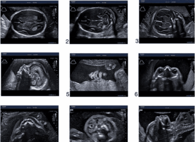
For a detailed anatomy scan,
a "2D" ultrasound is the primary tool used. This is because it provides highly accurate, precise measurements of internal organs and anatomical structures.
Therefore, 2D ultrasound is a vital diagnostic tool, not just a basic imaging technique.
4D Ultrasound (4-Dimensional Ultrasound)

Image Characteristics
Three-dimensional images with depth
Includes the dimension of time (allowing you to see the baby move in real-time)
What is it used for?
Provides a clear view of the baby's face
Allows you to see movements in real-time
Helps evaluate certain external physical structures, such as a cleft lip
So, is a 4D scan “necessary?”
No, it is not necessary for every pregnancy.
The most important medical assessment for your baby's health remains a detailed 2D anatomy scan.
A 4D ultrasound is a wonderful elective supplement that offers a clear external view and helps families build an early bond with their baby.

Comparison | 2D Ultrasound | 4D Ultrasound |
Image | Black and white, cross-sectional | Three-dimensional, live motion |
Primary Purpose | Structural evaluation | Viewing external appearance |
Medical Necessity | Essential | Optional (not medically required) |
Best Gestational Age | All trimesters | 24–32 weeks for the clearest facial views |
When is the best time to have these scans?
2D Scan: In every trimester, especially during weeks 18–22 for the anomaly scan
4D Scan: Between weeks 24–32 for the clearest views of the baby’s face
Ultrasound Safety
According to guidelines from the American College of Obstetricians and Gynecologists (ACOG) and the World Health Organization (WHO),
obstetric ultrasounds are safe when performed by qualified medical professionals for clinical indications.
Frequently Asked Questions
Is 4D more accurate than 2D?
It is not a matter of being more accurate; they simply serve different purposes.
2D ultrasounds are actually superior for detailed internal structural evaluations.
Why might the baby's face not be visible during a 4D scan?
The baby's position, the amount of amniotic fluid, and abdominal tissue thickness can all affect the image quality.
Is a 4D ultrasound needed at every visit?
No, it is not necessary and depends on the goal of that day's appointment.
Summary
If the goal is "a detailed health check of the baby" >> a 2D scan is the essential cornerstone.
If you want to see a clear picture of your baby's features >> a 4D scan can beautifully complement your experience.
At Femily Wellness Clinic, Ari,
all ultrasound scans are performed by OB/GYN specialists
who will guide you through the findings with detailed, easy-to-understand explanations at every step.
Written by
Dr. Wichadet Wichulada
OB/GYN Specialist, Maternal-Fetal Medicine (MFM) Fellow, King Chulalongkorn Memorial Hospital
Published researcher in the International Journal of Transgender Health