Home
Women’s Health Knowledge Center
2D vs. 4D Ultrasound: What’s the Difference? Which One Should You Get?
One of the questions pregnant mothers frequently ask is
“What’s the difference between 2D and 4D ultrasound?”
And does everyone need to have a 4D scan?
The truth is, both types serve different purposes
and they cannot be used interchangeably
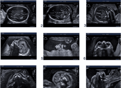
2D Ultrasound (2D Ultrasound)

Image characteristics
Black-and-white image
Seen as a cross-sectional image
What is it used for?
Estimate gestational age
Examine the baby’s organ structure
Check placental location and amniotic fluid
Screen for fetal abnormalities
Advantages
Medical standard
Allows detailed structural assessment
Necessary in every prenatal visit
In practice
In a detailed fetal anatomy scan (Anomaly scan)
what is primarily used in practice is “2D” because it provides accurate images for measuring organ size and examining anatomical structures (Anatomy)
Therefore, 2D is a primary medical tool, not just an ordinary device.
4D Ultrasound (4D Ultrasound)

Image characteristics
Three-dimensional image with depth
Adds the dimension of time (showing the baby moving in real time)
What is it used for?
See the baby’s face clearly
See movement in real time
Helps assess some external abnormalities, such as cleft lip
So, is 4D “necessary”?
It is not necessary for everyone
The most important part of checking the baby’s health remains a detailed structural assessment with 2D
4D is an additional tool that helps with viewing the baby’s external appearance and strengthening family bonding

Key point | 2D | 4D |
Image | Black-and-white, cross-sectional | Three-dimensional, with movement |
Main purpose | Assess structure | View external appearance |
Medical necessity | Necessary | Not necessary for every case |
Best gestational age | Any trimester | 24–32 weeks for the clearest facial view |
When is the best time to do it?
2D: Every trimester, especially 18–22 weeks (Anomaly scan)
4D: Around 24–32 weeks, the face is usually clearest
Ultrasound safety
According to the guidelines of American College of Obstetricians and Gynecologists and World Health Organization
obstetric ultrasound is considered safe when performed by medical professionals and used for appropriate indications
Frequently asked questions
Is 4D more accurate than 2D?
It is not about being more accurate, but about having different purposes
2D provides more detailed structural assessment
Why might the baby’s face not be visible during a 4D scan?
The baby’s position, amniotic fluid volume, and the mother’s abdominal fat can all affect this
Do I need to do it every time I come for an exam?
No, it depends on the purpose of the examination
Summary
If the goal is “a detailed check of the baby’s health” >> 2D is the key
If you want to clearly see your baby’s face >> 4D helps make that possible
At Femily Wellness Clinic, Ari
ultrasound examinations are performed by obstetrician-gynecologists
with detailed explanations at every step
Written by
Dr. Wichadet Vichchulada
Specialist in Obstetrics and Gynecology
Currently pursuing subspecialty training in Maternal-Fetal Medicine at King Chulalongkorn Memorial Hospital
Research published in the International Journal of Transgender Health